

Introduction
Ann Kimball and John W. Johnson Center for Cellular Therapeutics at Houston Methodist
Houston Methodist Dr. Mary and Ron Neal Cancer Center
The Food & Health Alliance within the Houston Methodist Lynda K. and David M. Underwood Center for Digestive Disorders, Immunology Center and the Fondren Inflammation Collaborative
Houston Methodist Cockrell Center for Advanced Therapeutics
Paula and Joseph C. “Rusty” Walter III
Translational Research Initiative
Jerold B. Katz Academy of Translational Research
Infectious Diseases Research Fund
George and Angelina Kostas Research Center for Cardiovascular Medicine
New Endowed Chairs Positions
EnMed
Center for Bioenergetics
result
Clinical Research
Outcomes, Quality and Healthcare Performances
Restorative Medicine
Precision Medicine
Science in Service
of
Medicineresult
President's letter
2022 Metrics
Cycle of Translation
Visionary Gifts of Hope


Introduction

Ann Kimball and John W. Johnson Center for Cellular Therapeutics at Houston Methodist

Houston Methodist Dr. Mary and Ron Neal Cancer Center

The Food & Health Alliance within the Houston Methodist Lynda K. and David M. Underwood Center for Digestive Disorders, Immunology Center and the Fondren Inflammation Collaborative

Houston Methodist Cockrell Center for Advanced Therapeutics

Paula and Joseph C. “Rusty” Walter III Translational Research Initiative

Jerold B. Katz Academy of Translational Research

Infectious Diseases Research Fund

George and Angelina Kostas Research Center for Cardiovascular Medicine

New Endowed Chairs Positions

EnMed

Center for Bioenergetics

From Discovery to Clinic


What is "Discovery to Clinic"?

Clinical Research


Houston Methodist Conducts First-Ever Study into a Challenging Situation

Can Regulating Cellular Aging Mitigate Both Cancer and Heart Disease?

Innovative Treatment for Chronic Rhinitis is Safe and Effective


Masters of Disguise: Glioblastomas Trick the Immune System by Masquerading as Reproductive Tissue
Improved Options for Patients with Severe Retinal Vascular Disease

A New FDA-Approved Treatment for Sufferers of Chronic Constipation

Houston Methodist joins the Gulf Coast Consortia

Outcomes, Quality and Healthcare Performance


New Findings on RNA Helicases May Yield New Intestinal Disease Therapy

Houston Methodist and Pennsylvania State University Collaborate on a Smartphone App That Could Revolutionize Stroke Diagnosis

New Frontiers to Improve Cardiovascular Medicine and Disease Management

Ongoing Lessons in a Pandemic

Transplants can Boost Survival Rate of Patients with Unresectable Liver Cancers

Telehealth Video Visits During the COVID-19 Pandemic – a Glimpse into the Future?

SARS-CoV-2 Induced Chronic Oxidative Stress and Endothelial Cell Inflammation May Increase Likelihood of Cardiovascular Diseases and Respiratory Failure

Restorative Medicine


Lessening Pain After Knee Replacement Surgery

Do Motor Neurons First Die in the Brain? Study Provides Clues about ALS Origins

Bringing Back Hand Function in People with Complete Spinal Cord Injury

Novel Vascular Engineering Platforms Are a Boon for Bioengineering

Ultra-high-Resolution Scanner Reveals if Knee Injury Advances to Osteoarthritis

Houston Methodist Model Demonstrates Reversal from Heart Failure State, Creating the Potential for Innovative Treatment Avenues

Precision Medicine


Rapidly Scalable, All-Inducible Neural Organoids Could Facilitate Drug Screening for Neurological Diseases

Importance of the Coronary Artery Calcium Score in Risk Assessment and Prevention of Atherosclerotic Cardiovascular Disease

COVID-19 Infection in Crucial Brain Regions May Lead To Accelerated Brain Aging

Interleukin 9 Secreting Polarized T Cells Show Potential in Solid and Liquid Tumor Treatment

The NanoLymph: Implantable. Adaptable. Anti-cancer


Discovery to Clinic
Ultra-high-Resolution Scanner Reveals if Knee Injury Advances to Osteoarthritis

Restorative Medicine
Ultra-high-Resolution Scanner Reveals if Knee Injury Advances to Osteoarthritis

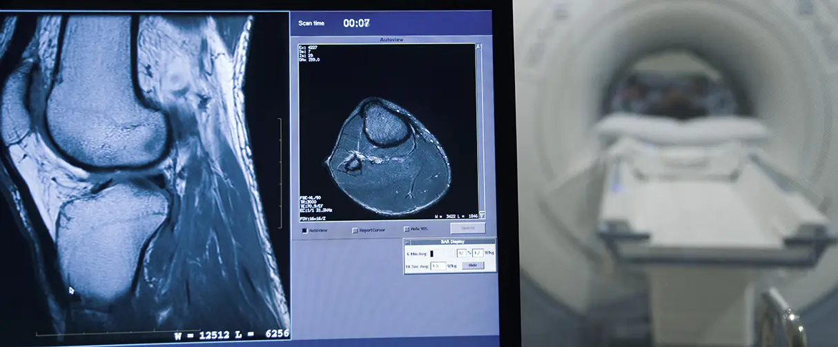
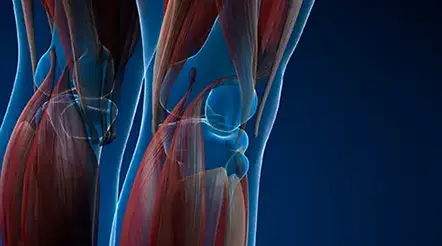
Capable of carrying up to five times an individual’s body weight, the knee joints are formidable powerlifters. However, when there is a severe injury to the joint, such as an anterior cruciate ligament (ACL) tear, the knee is vulnerable to slow recovery. In some cases, severe injury can set the knee on a path toward post-traumatic osteoarthritis, a long-standing painful condition that presently has no cure. To overcome this gap, Houston Methodist researchers are using ultra-high field 7 Tesla magnetic resonance imaging (MRI) with gadolinium dye as part of a protocol for tracking changes in the knee joints of rabbits after an experimentally induced ACL injury, a known yet understudied risk factor for osteoarthritis.
Although the study is in an animal model, the researchers said that ultra-high-resolution monitoring of the knee after an ACL tear sets the stage for identifying the root cause of osteoarthritis progression, a timeline for intervention and a potential platform to test the efficacy of therapeutic interventions.

Carly Filgueira, PhD
“What is available for treating people with osteoarthritis is pain management or in the worst case, a total knee replacement, and I thoroughly believe that we could do better,” said Carly Filgueira, PhD, Assistant Professor of Nanomedicine. “We're in the age where we now have the technology to identify if there could be a predisposition to osteoarthritis and prevent it from getting to the stage where you need to undergo an invasive surgical procedure.”
A report on the study is published in the journal Osteoarthritis and Cartilage Open.
Around 12% of all cases of osteoarthritis are caused by knee injuries. Further, the onset of symptoms of post-traumatic osteoarthritis can vary from a few months to years. During the early stages of recovery, when the patient is asymptomatic, biochemical and genetic changes occurring within the joint engender small morphological changes to the cartilage and bones within the knee. Some of these architectural modifications of the joint could contain telltale signs of future osteoarthritis, but they can be completely missed by routine radiographs that lack the resolution needed.
Flip the card over to learn more about post-traumatic osteoarthritis
However, MRI scans, particularly used with gadolinium dye, provide a much better view of the knee joint. This MRI technology, boosted with a 7 Tesla magnetic coil, has the power to yield an ultra-high-field view of the knee joint, particularly the cartilage. Further, the researchers noted that 7T gadolinium-enhanced MRI has never been used before to monitor the knee joint over a period of time to check for post-traumatic osteoarthritis progression.
“The big thing you have to think about is the thickness of the cartilage,” said Filgueira. “The 7T magnetic coil offers a high-resolution way to quantify very small-scale changes in cartilage thickness.”
Specifically, among other quantities, the researchers measured the delayed gadolinium-enhanced MRI of cartilage or the dGEMRIC index. Here, lower index values indicate degenerative changes or thinning in the articular cartilage. For their study, the team chose rabbits as their model systems and induced an ACL tear surgically in these animals. The large animal model allowed the researchers to reproduce the ACL tear across experiments and the ability to systematically follow the cartilage over 10 weeks of recovery. In addition, rabbit knees from the same animal, one with the ACL tear and one without, both fit into the coil of the 7T MRI scanner at the same time, facilitating comparisons between the joints.
Rotating 3D view of a rabbit knee joint taken with 7 Tesla MRI scanner. Dark yellow patches show bony outgrowths or osteophytes associated with the degeneration of the knee joint. Video provided by Carly Filgueira, PhD.
Upon refining their scanning protocol several times to optimize parameters that improved the signal to noise ratio, the team found that compared to the intact cartilage, there was a 35% and 39% decrease in the dGEMRIC index of the ACL on the medial side of the injured knee joint at seven and 10 weeks, respectively.
“This result is really key because we now have a time point when there is real evidence of cartilage degradation,” said Filgueira. “You can now say ‘OK, here's a time when we should be intervening’ and if we wait too long, any type of intervention is going to be too late because we've lost the cartilage.”
Thus, with an established model for post-traumatic osteoarthritis, the success of an intervention, such as nanomedicine, can be evaluated reliably. More broadly, their observations, although in rabbits, also make a strong case for early intervention and rehabilitation of the human knee after an ACL injury.
“Now, we have printed studies that show that you do need to intervene early, and you need to be put on a regimen that can prevent cartilage loss,” said Filgueira. “I think and hope that our study can help change people's mindsets.”
The research was supported by the Department of Orthopedics Pilot Project Initiative and funds from Houston Methodist Research Institute.
More from Discovery to Clinic


