Introduction
Ann Kimball and John W. Johnson Center for Cellular Therapeutics at Houston Methodist
Houston Methodist Dr. Mary and Ron Neal Cancer Center
The Food & Health Alliance within the Houston Methodist Lynda K. and David M. Underwood Center for Digestive Disorders, Immunology Center and the Fondren Inflammation Collaborative
Houston Methodist Cockrell Center for Advanced Therapeutics
Paula and Joseph C. “Rusty” Walter III
Translational Research Initiative
Jerold B. Katz Academy of Translational Research
Infectious Diseases Research Fund
George and Angelina Kostas Research Center for Cardiovascular Medicine
New Endowed Chairs Positions
EnMed
Center for Bioenergetics
result

Clinical Research
Outcomes, Quality and Healthcare Performances
Restorative Medicine
Precision Medicine

Science in Service
of
Medicineresult
President's letter
2022 Metrics
Cycle of Translation
Visionary Gifts of Hope
Introduction
Ann Kimball and John W. Johnson Center for Cellular Therapeutics at Houston Methodist
Houston Methodist Dr. Mary and Ron Neal Cancer Center
The Food & Health Alliance within the Houston Methodist Lynda K. and David M. Underwood Center for Digestive Disorders, Immunology Center and the Fondren Inflammation Collaborative
Houston Methodist Cockrell Center for Advanced Therapeutics
Paula and Joseph C. “Rusty” Walter III Translational Research Initiative
Jerold B. Katz Academy of Translational Research
Infectious Diseases Research Fund
George and Angelina Kostas Research Center for Cardiovascular Medicine
New Endowed Chairs Positions
EnMed
Center for Bioenergetics
From Discovery to Clinic
What is "Discovery to Clinic"?
Clinical Research
Houston Methodist Conducts First-Ever Study into a Challenging Situation
Can Regulating Cellular Aging Mitigate Both Cancer and Heart Disease?
Innovative Treatment for Chronic Rhinitis is Safe and Effective
Masters of Disguise: Glioblastomas Trick the Immune System by Masquerading as Reproductive Tissue
Improved Options for Patients with Severe Retinal Vascular Disease
A New FDA-Approved Treatment for Sufferers of Chronic Constipation
Houston Methodist joins the Gulf Coast Consortia
Outcomes, Quality and Healthcare Performance
New Findings on RNA Helicases May Yield New Intestinal Disease Therapy
Houston Methodist and Pennsylvania State University Collaborate on a Smartphone App That Could Revolutionize Stroke Diagnosis
New Frontiers to Improve Cardiovascular Medicine and Disease Management
Ongoing Lessons in a Pandemic
Transplants can Boost Survival Rate of Patients with Unresectable Liver Cancers
Telehealth Video Visits During the COVID-19 Pandemic – a Glimpse into the Future?
SARS-CoV-2 Induced Chronic Oxidative Stress and Endothelial Cell Inflammation May Increase Likelihood of Cardiovascular Diseases and Respiratory Failure
Restorative Medicine
Lessening Pain After Knee Replacement Surgery
Do Motor Neurons First Die in the Brain? Study Provides Clues about ALS Origins
Bringing Back Hand Function in People with Complete Spinal Cord Injury
Novel Vascular Engineering Platforms Are a Boon for Bioengineering
Ultra-high-Resolution Scanner Reveals if Knee Injury Advances to Osteoarthritis
Houston Methodist Model Demonstrates Reversal from Heart Failure State, Creating the Potential for Innovative Treatment Avenues
Precision Medicine
Rapidly Scalable, All-Inducible Neural Organoids Could Facilitate Drug Screening for Neurological Diseases
Importance of the Coronary Artery Calcium Score in Risk Assessment and Prevention of Atherosclerotic Cardiovascular Disease
COVID-19 Infection in Crucial Brain Regions May Lead To Accelerated Brain Aging
Interleukin 9 Secreting Polarized T Cells Show Potential in Solid and Liquid Tumor Treatment
The NanoLymph: Implantable. Adaptable. Anti-cancer


Discovery to Clinic
Restorative Medicine
Do Motor Neurons First Die in the Brain? Study Provides Clues about ALS Origins
Do Motor Neurons First Die in the Brain? Study Provides Clues about ALS Origins

Share this story


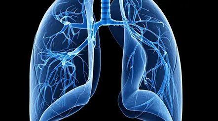
Theoretical astrophysicist Stephen Hawking is known as much for his revolutionary theories on black holes as for the motor neuron disease that left him crippled for most of his adult life. Although amyotrophic lateral sclerosis (ALS) eventually causes a complete loss of muscle control, its rate of progression can vary significantly between patients, making the tracking of the disease an important clinical need.
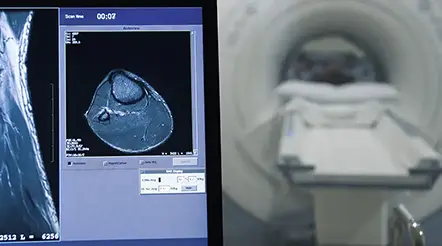
In a Journal of Neurology study, Houston Methodist researchers evaluated the feasibility of the wearable transcranial rotating permanent magnet stimulator (TRPMS) as a prognostic tool for ALS.

Santosh Helekar, MD, PhD
“Our transcranial device is much more sensitive and involves milder stimuli than the conventional transcranial magnetic stimulators and has multiple therapeutic applications, including in stroke and depression,” said Santosh Helekar, MD, PhD, Associate Professor of Neurosurgery. “In this study, we tested if the device could be used to track the progression of ALS over a period of time.”
Specifically, using the TRPMS, the researchers were able to distinguish between two competing theories of the origins of the disease.
ALS begins as weakness and stiffness in the muscles, eventually affecting all voluntary muscles in the body, causing difficulty in speaking, moving and breathing. About 10% of the people with ALS have an underlying genetic cause, such as a mutation in the enzyme superoxide dismutase. However, in most cases, ALS occurs spontaneously in people with no family history of the disease.
The disease is marked by the death of motor neurons in both the central and peripheral nervous systems. In a clinical setting, a transcranial magnetic stimulator is used to probe the health of cortical motor neurons. When the noninvasive device is placed on the head and switched on, it generates strong magnetic fields that stimulate the motor cortex. This induced cortical activity in turn evokes electric signals, called motor-evoked potentials, in the muscles. Thus, a change in the amplitude of the motor-evoked potential reflects a change in the activity of the motor cortex; that is, lower activity would indicate that neurons have died due to the disease.
Previous studies using transcranial stimulation have shown that during the early stages of ALS, cortical motor neurons let out a final gasp of increased electrical activity before dying. This hyperexcitability causes a commensurate increase in the intensity of motor-evoked potentials in the muscles of the hand. Although useful in disease prognosis, transcranial magnetic stimulation is not always a reliable indicator of the extent of cortical motor neuron damage.
“The motor-evoked potential amplitude even in normal subjects can be very variable,” said Helekar. “The same stimulation conducted multiple times can give different results, so we wanted to see if we can get consistent results with our device, which uses weaker but more focused stimulation.”
Unlike the conventional transcranial magnetic stimulator that uses a single large electromagnet, Helekar’s TRPMS uses smaller permanent magnets that are embedded within a wearable cap. These magnets are positioned in different locations on the head and can stimulate specific brain regions when spun with battery-driven electric motors. Thus, the device produces weaker magnetic fields at specific locations in the brain that then drive spontaneous motor unit potentials in muscles.
When the researchers analyzed cross-sectional and longitudinal electromyographic data from 40 and 20 ALS patients, respectively, who received TRPMS stimulation, they found that for some patients, the counts of the motor unit potentials in the hand muscles changed with ALS progression in an inverted V shape. In other words, in these people, the hyperexcitability of the cortex occurred in the intermediate rather than in the beginning stage of the disease.
There are two competing ideas for the origin of ALS: The dying forward hypothesis where the cortical neurons die first and the peripheral motor neurons follow, and then there is the dying back hypothesis where the pathology starts in the periphery,” said Helekar. “But we saw a mix in our patient pool — most appear to have the dying back type of pathogenesis but some have the dying forward kind and then others might have a combination.”
The researchers noted that their study provides a line of evidence to distinguish between the two hypotheses for ALS pathogenesis. Thus, their results need to be cross validated with other scientific approaches and with more patients for more conclusive validation. However, despite this limitation, Helekar said that TRPMS is an important tool to track ALS disease progression.
“The progression of motor deficits can occur really rapidly or slowly. Typically, patients die of ALS in 3-4 years, but some, like Stephen Hawking, can live for 50 years,” said Helekar. “Since TRPMS allows us to track the progression of the disease, we now have a tool to potentially check if any disease-modifying treatment is working.”
The study was funded in part by a grant from the Houston Methodist Specialty Physicians Group.
More from Discovery to Clinic