

Precision Medicine
Interleukin 9 secreting polarized T cells show potential in solid and liquid tumor treatment

Chimeric antigen receptor (CAR) T cell immunotherapy is useful for various malignancies. However, the biggest hurdle is the relapse seen in most patients. A relapse can even be seen in patients with complete remission during early treatment stages. There is a pressing need to enhance the therapeutic efficacy of CAR-T cells to achieve long-term remissions. Antigen loss and poor persistence of infused CAR-T cells are behind the paucity of success of CAR-T cell immunotherapy. The latter is due to CAR-T cell apoptosis and T cell exhaustion - an intricate phenomenon dependent upon patient age, starting state of T cells and the tumor microenvironment.
Antigen-specific interleukin 9 (IL-9) secreting (CD4+ Th9 or CD8+Tc9) T cells are a sub-category of T cells demonstrated to have greater anti-tumor efficacy in mouse models relative to Th1, Th17, or Tc1/CTL T cells. Compared to other T cell subsets, these T cells express lower levels of cytolytic proteins and exhaustion markers and distinct cytokine profiles. Based on this premise, Qing Yi, MD, PhD, professor of oncology at the Houston Methodist Cancer Center, hypothesized that human CAR-T cells polarized ex vivo under Th9 culture conditions (T9 CAR-T cells) are more potent in destroying tumors in vivo relative to CAR-T cells polarized under standard Th1/Tc1 culture conditions (T1 CAR-T cells). By polarizing and expanding CD19 or GPC3 CAR-T cells under Th9 culture conditions, Yi created IL-9 secreting CAR-T cells with a superior anti-tumor potency against CD-19 expressing acute lymphoblastic leukemia and GPC3 expressing liver carcinoma.
T9 cells secrete high IL-9 levels and low interleukin-gamma (IL-ɣ) levels, whereas the converse is true of T1 cells. A series of analyses were conducted to compare T9 CAR-T cells with T1 CAR-T cells in terms of anti-tumor activity, proliferative capacity, and persistence within the tumor microenvironment.
Cytotoxicity assays tested the tumor-killing ability of T9 CAR-T cells and revealed a higher cytolytic potency of T9 cells, relative to T1 CAR-T cells. To unravel the mechanisms behind this, gene set enrichment analysis (GSEA) was performed to evaluate expression levels of genes associated with apoptosis, cell cycle and proliferation. T1 CAR-T cells were found to be enriched in apoptosis genes, whereas the T9 cells had a more robust expression of genes associated with G2/M transition, DNA replication and cell cycle regulation. This suggested greater proliferative capacity in T9 cells. To evaluate this further, T9 CAR-T cells were exposed to CD-19 expressing tumor cells and found to have a five-fold higher proliferative capacity in culture relative to T1 CAR-T cells. Moreover, T9 CAR-T cells demonstrated reduced apoptosis compared to T1 CAR-T cells.
A comprehensive analysis of the T9 transcriptome demonstrated distinct cytokine expression profiles and reduced T cell exhaustion and differentiation markers. This result aligned with mouse model studies and suggested functional differences between these two CAR-T cell types. Further, GSEA indicated that the human T9 CAR-T cells were enriched in the central memory stage, whereas the human T1 CAR-T cells in the effector memory stage.
To understand the differentiation states of CAR-T cells, Yi performed GSEA of RNA sequencing data which affirmed that T9 cells had enhanced expression of genes significant for central memory T cells (Tcm), whereas T1 cells had higher expression of genes important for effector memory T cell (Tem). Relative to T1 CAR-T cells, T9 cells have diminished expression of genes controlling effector differentiation including PU.1, eomesodermin, T-box 21 and interferon regulatory factor-1.
Compared to T1, T9 CAR-T cells are less terminally differentiated and exhausted. Flow cytometry revealed that T9 cells constituted a higher proportion of Tcm and T1 CAR-T cells a higher proportion of Tem. Tcm and Tem cell subsets have unique transcriptional profiles and granzyme production- with Tcm exhibiting a significantly advanced respiratory capacity.
CAR-T cell immunotherapy hasn’t been terribly successful in solid tumors owing to poor migration and infiltration of CAR-T cells into tumors. In addition to in vitro assays, Yi tested the capacity of human T9 CAR-T cells to repress solid tumor growth in murine tumor models. Tumor-bearing mice treated with T9 CAR-T cells showed greater T9 CAR-T cell infiltration into tumor sites, greater survival and reduced tumor growth, as compared to T1 CAR-T treated mice. Thus, T9 CAR-T cells show considerable therapeutic efficacy and potential for solid human tumors.

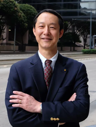
Qing Yi, MD, PhD
Our findings highlight a promising clinical potential of human IL-9 secreting T cells for CAR-T cell immunotherapy for human cancers, attributed to their central memory phenotype and that they are less exhausted, hyperproliferative and long-lived in vivo. Furthermore, these CAR-T cells exerted a greater anti-tumor efficacy against both liquid and solid tumors in human xenografted mouse models in comparison with classically polarized, IFN-ɣ secreting CAR-T cells. These unique features of T9 CAR-T cells endow them with greater potential for clinical immunotherapy, which can lead to increased frequency of complete remission, high survival rates, and decreased number of patient relapse after treatment.

Qing Yi, MD, PhD, Professor of Oncology
Houston Methodist Cancer Center
Treatment outcomes in solid and liquid tumor patients can be significantly enhanced by harnessing the hyperproliferative capacity (both in vitro and in vivo) and robust antitumor potency of human T9 CAR-T cells. Engineering T cells with a CAR is justifiable and human T9 CAR-T cell therapy is likely to lead to better long-term remission amongst cancer patients.
Lintao Liu, Enguang Bi, Xingzhe Ma, Wei Xiong, Jianfei Qian, Lingqun Ye, Pan Su, Qiang Wang, Liuling Xiao, Maojie Yang, Yong Lu, Qing Yi. Enhanced CAR-T activity against established tumors by polarizing human T cells to secrete interleukin-9. Nature Communications (2020) 11: 5902. doi: 10.1038/s41467-020-19672-2
This work was supported by NCI R01 CA200539 and Cancer Prevention & Research Institute of Texas Recruitment of Established Investigator Award (RR180044). Q.Y. and his research group are also supported by NCI R01s (CA211073, CA214811, and CA239255).
Abanti Chattopadhyay, PhD, July 2021
Related Articles


