

Precision Medicine
New high-throughput analytic enables differentiation of tumor cells within its microenvironment

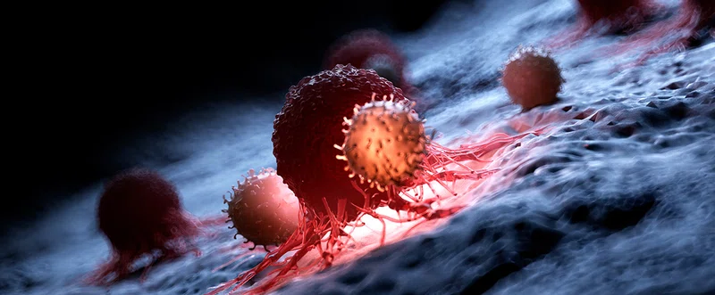
Illustration of white blood cells attacking a cancer cell
One of the paramount goals in the treatment and eradication of cancer is understanding what causes cancer. To this end, it is crucial to be able to study tumor cells, their genetics, and epigenetics in isolation. Houston Methodist experts from the Center for Bioinformatics and Computational Biology have devised a new technology that enables clinicians to distinguish the tumor cells from among the non-cancerous components of the microenvironment, which may lead to more informed cancer treatment approaches.
Rather than existing in isolation, tumor cells are always part of the local microenvironment along with stromal cells, immune cells, and components of the extracellular matrix. The ability to discern tumor cells from normal cells within the tumor microenvironment and to delineate the clonal substructure within tumors has been a challenge in the field. Various methodologies are available that aim to study human tumors by analyzing single-cell transcriptomes. The ability to estimate genomic copy numbers from RNA read counts has been demonstrated previously by techniques such as inferCNV and HoneyBadger. However, these are equipped to only analyze data from low throughput single-cell RNA sequencing methods such as Fluidigm and SMART-seq2.
Recent breakthroughs in transcriptomic technologies allow high throughput-sequencing with the generation of single-cell transcriptomic data and measurement of gene expression in thousands of singular cells simultaneously. These high-throughput sequencing technologies such as Drop-Seq, Indrop, 10X Chromium (microdroplet systems) and Wafergen iCELL8, SeqWell, CelSee (nanowells) provide a major cost advantage by allowing sequencing of thousands of individual cells simultaneously for less than 1 USD per cell.
Aneuploidy is prevalent in 88% of human tumors. In contrast, non-tumorous stromal and immune cells in the tumor microenvironment do not display aneuploidy or copy number variation. Using CopyKAT analysis, Gao successfully distinguished tumor cells from normal cells in 21 different tumors with 98% accuracy. These different tumor types included triple-negative breast cancer, pancreatic ductal adenocarcinoma, anaplastic thyroid cancer, invasive ductal carcinoma, and glioblastoma, which suggests that CopyKAT can be useful in distinguishing tumor cells from normal cells in myriad solid human tumors. Furthermore, the CopyKAT technique successfully inferred clonal subpopulations by detecting variations in gene expression profiles of cancer genes, markers of epithelial-to-mesenchymal transition, DNA repair, apoptosis, and hypoxia.
In a research study led by Ruli Gao, PhD, assistant professor of cardiovascular sciences from the Houston Methodist Center for Bioinformatics and Computational Biology, the phenomenon of aneuploidy in tumor cells was leveraged to develop an integrative Bayesian segmentation approach called CopyKAT to estimate genomic copy number profiles in high-throughput single-cell RNA sequencing data. CopyKAT, which stands for copy number karyotyping of aneuploid tumors, offers practical applications of identifying tumorous cells of a variety of solid human tumor types as well as an understanding of the clonal substructures in the tumor microenvironment. This study was performed in collaboration with Nicholas E. Navin, PhD, associate professor in the Department of Bioinformatics and Genetics at the MD Anderson Cancer Center.

Ruli Gao, PhD,
assistant professor of cardiovascular sciences from the Houston Methodist Center for Bioinformatics and Computational Biology
These studies will greatly improve understanding of the malignant gene expression programs of tumor cells by providing a pure tumor cell signal, whereas previous bulk RNA-seq methods have been challenged by the intermixing of stromal and immune cells with the tumor cells, resulting in incorrect assessment of cancer phenotypes. Moreover, these studies will lead to new insights into how chromosome alterations lead to gene dosage effects that reprogram cancer phenotypes in human tumors during disease progression.

Ruli Gao, PhD and Nicholas E. Navin, PhD
Gao is Assistant Professor of Cardiovascular Sciences from the Houston Methodist Center for Bioinformatics and Computational Biology. Navin is associate professor in the Department of Bioinformatics and Genetics at the MD Anderson Cancer Center
Yet, CopyKAT has some notable limitations. Firstly, it cannot be applied to all tumor types since not all cancers show considerable copy number variations. Exceptions include pediatric and hematopoietic cancers such as acute myeloid leukemia and chronic lymphocyte leukemia. Secondly, CopyKAT analysis cannot detect copy number aberrations stemming from genomic events such as chromosomal structural rearrangements, indels, and somatic mutations. Its application is limited to detecting changes in read depth. Thirdly, due to variability of the 3’ end single-cell RNA sequencing data, CopyKAT analysis cannot provide information on the genomic copy number of individual cells with unique genotypes. Taken together, CopyKAT is a technique useful in deciphering and providing analysis of subclones within a significant range of tumor masses.
Correct assessment of cancer phenotypes and correlation between subclone gene expressions and cancerous phenotypes is crucial in understanding how cancer is caused at the molecular level as well as the mechanisms of cancer progression. CopyKAT is thus a potentially valuable tool in identifying tumor cells in high-throughput, single-cell RNA sequencing experiments that generate limited data and has wide applications in solid human tumors and efficient therapeutics.
Ruli Gao, Shanshan Bai, Ying C Henderson, Yiyun Lin, Aislyn Schalck, Yun Yan, Tapsi Kumar, Min Hu, Emi Sei, Alexander Davis, Fang Wang, Simona F. Shaitelman, Jennifer Rui Wang, Ken Chen, Stacy Moulder, Stephen Y. Lai and Nicholas E. Navin. Delineating copy number and clonal substructure in human tumors from single-cell transcriptomes. Nature Biotechnology May 2021, 39(5):599-608.
This research study led by Ruli Gao, PhD was commenced at MD Anderson Cancer Center during her Postdoctoral Fellowship with Nicholas E. Navin, PhD and completed at Houston Methodist where she is currently appointed as assistant professor.
Abanti Chattopadhyay, PhD, June 2021


