result
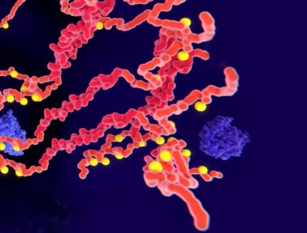
Precision medicine
Urgent Need for Improved Vascular Imaging Training Programs
Urgent Need for Improved Vascular Imaging Training Programs
Improving Cardiovascular Disease Management Through Enhancing Training Opportunities in Vascular Imaging
Improving Cardiovascular Disease Management Through Enhancing Training Opportunities in Vascular Imaging
Share this story


With the increase in rates of diabetes and obesity and the concomitant increase in mortality due to cardiovascular diseases, there is a critical need for vascular surgery services in both planned and unplanned clinical settings. In a review study published in the Methodist DeBakey Cardiovascular Journal, Trisha Roy, MD, PhD, clinical scholar and assistant professor of Cardiovascular Surgery at Houston Methodist has shed light on the gaps that currently exist in the training regimens of vascular surgery and how advances in medical imaging are reshaping the practice of vascular surgery. Insights drawn from this review study can help in creating better training programs on advanced vascular imaging techniques which may lead to improvements in patients’ recovery times, disease outcomes and overall quality of life.

The ability to understand and interpret imaging is critical to treating patients with vascular disease. Cardiology has shown how dedicated training in advanced imaging has had a synergistic effect of giving cardiologists more information to treat patients while also improving the imaging modalities themselves. As an end-user of imaging techniques, the perspectives of cardiologists combined with imaging training have led to major advances in cardiac MRI, coronary computed tomography, positron emission tomography perfusion imaging, and more. Improved diagnostics propelled the cardiology specialty as a whole as clinicians developed a new understanding of the structure and function of the heart and vessels. By comparison, vascular surgery has not seen similar growth in imaging techniques, and the lack of training has limited the specialty as a whole from integrating and developing new imaging modalities into clinical practice.

Trisha Roy, MD, PhD
Assistant professor of Cardiovascular Surgery
at Houston Methodist

Trisha Roy, MD, PhD
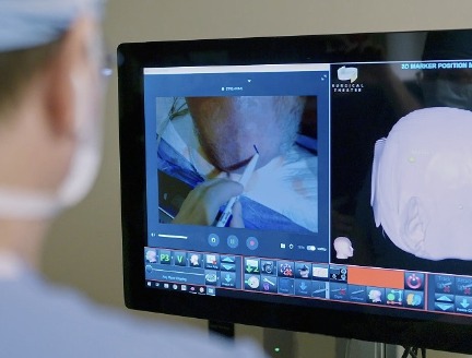
There are many benefits of minimally invasive surgery, including less post-operative pain, fewer complications, less scarring and a faster recovery. However, the successful implementation of minimally invasive procedures depends on imaging modalities and the vascular surgeon’s comfort in using advanced imaging modalities.
Imaging modalities and techniques have grown exponentially in the fields of interventional radiology, interventional and diagnostic cardiology and neuroradiology. On the other hand, duplex ultrasound, digital subtraction angiography and computer tomography (the three most commonly used vascular surgery techniques) have had limited advancements in the past 20 years. Notably, vascular surgery training programs currently do not offer formal training in vascular imaging other than duplex ultrasound, which involves using two modes of ultrasound waves to study blood flow speed and the structure of the leg veins.

Heart surgeons and operating room nurse during a heart valve operation. Getty Images.
Furthermore, vascular surgeons receive fragmentary training in magnetic resonance imaging (MRI), which leads to a lack of confidence in interpreting MRI thereby restricting its clinical usage. This instance underscores the gap in the knowledge and training of vascular surgeons that needs to be rectified.
According to Roy, “The ability to understand and interpret imaging is critical to treating patients with vascular disease. Cardiology has shown how dedicated training in advanced imaging has had a synergistic effect of giving cardiologists more information to treat patients while also improving the imaging modalities themselves. As an end-user of imaging techniques, the perspectives of cardiologists combined with imaging training have led to major advances in cardiac MRI, coronary computed tomography, positron emission tomography perfusion imaging, and more. Improved diagnostics propelled the cardiology specialty as a whole as clinicians developed a new understanding of the structure and function of the heart and vessels. By comparison, vascular surgery has not seen similar growth in imaging techniques, and the lack of training has limited the specialty as a whole from integrating and developing new imaging modalities into clinical practice.”
There is certainly a need for systematic training on vascular imaging techniques as various vascular conditions are on the rise. A case in point is peripheral arterial disease (PAD), which caused 74,000 deaths in 2019 with 10 million new PAD cases. Furthermore, nearly 113 million people lived with PAD in 2019, which is a very painful condition entailing leg numbness and weakness, coldness in the lower leg and stiffness in the hips.
For improved management of patients with cardiovascular disease, vascular surgeons must receive thorough training in the state of the art imaging techniques. Vascular imaging techniques play crucial roles in aiding clinicians to make informed decisions regarding planning procedures. Thorough knowledge of key vascular imaging techniques such as vascular MRI can help clinicians not only evaluate the patients’ vasculature but also develop novel imaging techniques that can push the envelope toward superior patient diagnosis and care.
Kavya Sinha, Marton Berczeli, Alan B Lumsden, Trisha L Roy. Imaging: New Frontiers in Vascular Training. Methodist Debakey Cardiovasc J. 2022 Jun 3;18(3):39-48. doi: 10.14797/mdcvj.1093.
Abanti Chattopadhyay, PhD
June 2023
Share this story