

Restorative medicine
Improved Translational Models to Probe Brain Hemorrhage

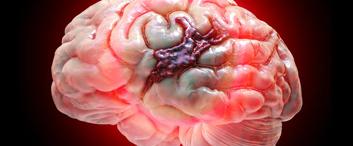
A battery of validated rodent behavioral tests now facilitates investigating the underpinnings of neurocognitive deficits after a subarachnoid hemorrhage.
In a recent study, researchers at the Houston Methodist Neurological Institute have validated a series of behavioral tests to establish a rodent model system for studying the aftermath of subarachnoid hemorrhage. These tests, they said, can facilitate future investigations into the basis of learning and memory deficits due to the catastrophic hemorrhage and ways to evaluate the efficacy of therapeutic interventions.
“In this research, we examined sophisticated behavioral tests in mice to establish a robust baseline to investigate neurological deterioration after a subarachnoid hemorrhage,” said Gavin Britz, MBBCh, Candy and Tom Knudson Distinguished Centennial Chair in Neurosurgery and professor in the Department of Neurosurgery. “This baseline is particularly important as we develop therapeutics, such as devices and drugs, for treating the devastating effects of hemorrhagic strokes.”
While the least frequent type of stroke, subarachnoid hemorrhages have a fatality of 40% and are often precipitated by a ruptured aneurysm. In the early days after the hemorrhage, injuries are largely due to intracranial pressure, which causes a decrease in cerebral blood flow and a wave-like spread of depressed electrical activity throughout the brain. Between 3–14 days after the subarachnoid hemorrhage, around one-third of patients experience a worsening of neurological symptoms, such as a fluctuating decline in consciousness. Overall, 95% of the patients are left with permanent neurological, psychological and cognitive deficits.
Brain areas, particularly those that are related to learning and memory, have been strongly implicated in the neurocognitive decline post subarachnoid hemorrhage. For example, some patients have been reported to have atrophy in the hippocampus, a region that plays a vital role in making and storing memories. However, the mechanisms that underlie hippocampi volume loss after a subarachnoid hemorrhage have yet to be fully revealed.

This research will also allow explorations into different possible mechanisms, including that of the complement pathway of the immune system, spinal fluid flow, glymphatic flow, that can all contribute to the neurocognitive deficits observed in patients after a stroke.


Gavin W. Britz, MBBCh
Candy and Tom Knudson Distinguished Centennial Chair in Neurosurgery, Houston Methodist
In that regard, due to their genetic tractability, rodents are a powerful model system to investigate questions on the impact of subarachnoid hemorrhage on neural tissue. But currently, there are no prescribed behavioral tests to evaluate the short-and long-term neurocognitive deficits in these animal models. To fill this gap, Britz and his team evaluated a series of neurocognitive tests to probe the loss of sensory, motor and cognitive function in mice after a subarachnoid hemorrhage.
Before these tests, the researchers evoked an experimental subarachnoid hemorrhage in mice by rupturing arteries in the Circle of Willis region located at the base of the brain. Next, they made the animals perform a series of tests at different time points after the induced hemorrhage. Some of the tests included motor and sensory tests, an open field test for anxiety, the Y and Barnes maze for spatial learning, an object recognition test and a social interaction test.
When the team compared the results of the behavioral tests between mice that received the subarachnoid hemorrhage with those that did not, they found that the experimental mice displayed impairments in spatial memory and social interaction. In a month after the hemorrhage, the experimental mice developed anxiety-like behavior and showed further deterioration of spatial learning and memory, reminiscent of delayed neurological deficits in humans.
The results of the behavior studies are published in Translational Stroke Research.
Britz noted that although their current study included a whole battery of behavioral tests, in the long term, the team plans to refine the number of tests needed to evaluate subarachnoid hemorrhage-induced neurocognitive deficits. Further, these tests may be used to assess the efficacy of drugs in mitigating neurocognitive symptoms post hemorrhage.
“Now we can objectively test if a therapeutic measure will make a difference,” said Britz. “Not just that, this research will also allow explorations into different possible mechanisms, including that of the complement pathway of the immune system, spinal fluid flow, glymphatic flow, that can all contribute to the neurocognitive deficits observed in patients after a stroke.”
Regnier-Golanov, A.S., Gulinello, M., Hernandez, M.S. et al. Subarachnoid Hemorrhage Induces Sub-acute and Early Chronic Impairment in Learning and Memory in Mice. Transl. Stroke Res. (2022). https://doi.org/10.1007/s12975-022-00987-9
Vandana Suresh, PhD, June 2022
Related Articles

RESTORATIVE MEDICINE
Lessening Pain After Knee Replacement Surgery
Infusing morphine and vancomycin directly into the tibia during knee replacement reduces pain and pain medication needed after surgery.

RESTORATIVE MEDICINE
Novel Vascular Engineering Platforms Are a Boon for Bioengineering
Owing to a dearth of donated organs for transplantation, scientists at Houston Methodist have made a significant breakthrough the field of organ engineering


