


result
clinical research
Personalizing Care for Peripheral Arterial Disease Patients
Personalizing Care for Peripheral Arterial Disease Patients
Peripheral arterial disease (PAD) is a
chronic, circulatory condition that affects
more than 236 million people worldwide.
Peripheral arterial disease (PAD) is a
chronic, circulatory condition that affects
more than 236 million people worldwide.
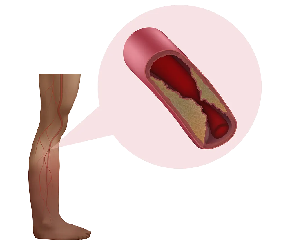
According to the Centers for Disease Control and Prevention, approximately 6.5 million people in the United States aged 40 and older have PAD, which is caused by an atherosclerotic plaque build-up inside arteries that leads to a blood-flow blockage to the limbs. It is more common in legs than arms, and risk factors include smoking, hypertension, diabetes, atherosclerosis, and age (older than 60 years).
Share this story
PAD increases the risk of stroke, cardiovascular death, myocardial infarction, coronary artery disease and cerebrovascular disease. PAD management includes pharmacological treatments and revascularization of blood vessels. Percutaneous vascular intervention (PVI)—a minimally invasive procedure is attempted to unblock occluded arteries. In cases of impenetrable plaques, PVI has led to worsened secondary bypass outcomes and amputation.

Trisha Roy, BASc, MD,
PhD, FRCSC
Trisha Roy, BASc, MD, PhD, FRCSC, Assistant Professor of Cardiovascular Surgery, published a case report in the Methodist DeBakey Cardiovascular Journal 2023 to illustrate the importance of Magnetic Resonance Imaging (MRI) histology in PVI planning to minimize unwanted patient outcomes. This report discusses the case of a 64-year old female patient with critical limb ischemia (CLI) who underwent an above-knee amputation because of a failed PVI attempt to recanalize the limb.
According to the American College of Cardiology/American Heart Association Practice Guidelines, there are four types of PAD presentation: asymptomatic, claudication, CLI and acute limb ischemia. CLI is a chronic form of PAD that entails severe blockage of arteries in the legs with reduced blood flow. The main CLI symptom is severe pain in the feet and legs even when resting is referred to as ischemic rest pain. Other symptoms include numbness in the legs, gangrenes, diminished pulse, skin infections and ulcers.
PAD symptoms are related to the blood flow being restricted due to blockages inside the arteries. Blockages result from the build-up of fatty deposits or plaque and can be hard (calcium and dense collagen) or soft. Appropriate interventional strategies for blood flow restoration are critical to prevent amputation and death.
The 64-year-old CLI patient had a history of diabetes, hyperlipidemia, hypertension, coronary artery disease and atrial fibrillation. The patient’s right foot had worsening rest pain, non-healing wounds and ulcers. This patient had an impenetrable plaque in her popliteal artery. Failed PVI attempts were followed by failed bypass attempts, eventually requiring an above-knee amputation. Typically, CLI patients have a 20% risk of losing a leg within a year and a 20% risk of dying (usually from a heart attack). According to the Amputee Coalition, about 185,000 amputations occur in the United States each year. Nearly 54% of limb loss cases are due to diabetes and PAD.
Using ultrashort echo time (UTE) and T2-weighted (T2W) sequences, MRI histology can be used to determine if blockages are hard or soft. This offers a good predictive value of not only identifying patients who are at a high risk of failing the endovascular intervention but also predicting whether their lesions would require more adjunctive devices.
Nearly 54% of limb loss cases are due to diabetes and PAD
Atheromatous plaques composed of dense collagen are missed on preoperative imaging. Consequent decisions to perform percutaneous vascular intervention often result in failure as dense collagen lesions are difficult to penetrate with guidewire. MRI histology can distinguish between soft plaque and hard plaque composed of calcium and dense collagen. This novel imaging technique offers a comparative edge in selecting patients suited better for percutaneous vascular intervention versus open surgery in advance.
Trisha Roy, BASc, MD, PhD, FRCSC
Assistant Professor of Cardiovascular Surgery
There are a couple of anatomic scores for PAD that indicate if a lesion or a patient will be better served by endovascular treatment or an open bypass. These scores are based on the length and degree of stenosis. While the aforementioned do not provide any details about what the occlusion is made of. Soft lesions (whether long or short) can be recanalized using endovascular procedures whereas hard lesions are impenetrable.
“As identified in this case, atheromatous plaques composed of dense collagen are missed on preoperative imaging. Consequent decisions to perform PVI often result in failure as dense collagen lesions are difficult to penetrate with guidewire. MRI histology with UTE and T2W sequences can distinguish between soft plaque and hard plaque composed of calcium and dense collagen. This novel imaging technique offers a comparative edge in detecting PVI failures and selecting patients suited better for PVI versus open surgery in advance,” Roy said.
With the advent of new sophisticated MRI scanners, researchers now have access to refined methods because of the availability of a wide array of sequences (in addition to UTE and T2W) and material equipment.
Roy added, “PAD is a real epidemic, and it is growing along with diabetes and renal failure. Our understanding of PAD is quite poor and the imaging techniques that we use have been the same for decades. So, having the ability to be advanced with how we're diagnosing PAD, and tailoring our treatment can help patient outcomes, particularly because there's a big spectrum to what the blockages can be made of. MRI histology is a good tool to shed light on what these blockages are composed of and thereby predict endovascular failure. Right now, our outcomes are quite poor and we're having to intervene on these patients again and again, each time more futile than the last. We hope that by being able to image and understand what we're treating, we have a better understanding of how to treat individual patients so that they have the best possible outcomes.”
Shah KJ, Benfor B, Karmonik C, Lumsden AB, and Roy, TL. To Cross or Not to Cross: Using MRI-Histology to Characterize Dense Collagenous Plaque in Critical Limb Ischemia. Methodist Debakey Cardiovasc J. 2023; 19(1): 1–6. doi: 10.14797/mdcvj.1186
This work was supported by the Jerold B. Katz Foundation (Katz Academy of Translational Research).
Abanti Chattopadhyay, PhD
April 2024
Related Articles

Share this story