
Share


result
Clinical Research
Houston Methodist researchers review the similarities and contrasts of amphibian regeneration and mammalian cancer from an evolutionary biology perspective

Humans have known for a time that certain salamanders can regenerate their limbs after losing one. Though in most cases this happens spontaneously, research has shown that if cells from a forelimb are placed where a lost hind limb was, the cells can grow a forelimb in place of a hind limb.
The ability to regenerate is present across a range of species and may have evolved as a strategy for escaping predators.
It appears that an axolotl (walking fish) must first have the ability to grow said limbs in the embryotic state in order to have the ability to regenerate the limb, but once it has that ability, the sky seems to be the limit regarding how many times these amphibians can regenerate limbs, eyes, jaws, tails and even its heart after injury. Where the limb is lost seems to not matter – endogenous cells appear to be able to regenerate as needed.
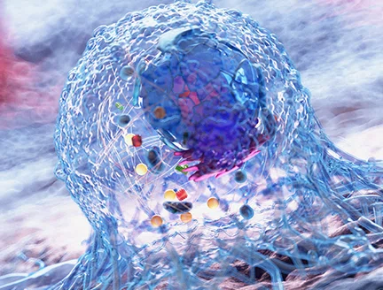
Houston Methodist investigators recently reviewed the link between regeneration capacity and tumor suppression. They compared mammal embryos to adults with highly regenerative vertebrates. In amphibians (essentially newt sand salamanders), the reparative process relies on a precise molecular and cellular machinery capable of sensing abnormal signals and actively reprograming or eliminating them. The tumor also retains common functional attributes, and the immune system plays a pivotal role in maintaining a physiological balance to provide surveillance against tumor initiation or to support its initiation and progression. Investigators speculated that susceptibility to cancer development in adult mammals may be determined by the loss of an advanced regenerative capability during evolution and believe that gaining mechanistic insights into how regenerative capacity linked to tumor suppression is postnatally lost in mammals might illuminate an as yet unrecognized route to cancer treatment.

Bruna Corradetti, Ph.D.
Assistant Research Professor of Nanomedicine
Regeneration requires instructive signals to regulate cells, which results in a finite number of cells that divide, differentiate and repair. Once repair is complete, these cells stop the process. The process is not always perfect and abnormal cell growth can lead to a malignant transformation. Researchers are not quite sure why that happens but hypothesize that the formation of these tumors might be dependent on impaired or incomplete regeneration.
This hypothesis is supported by experimental evidence illustrating that during local mammalian tissue repair, chronic exposure to damage and subsequent attempted repairs may induce an accumulation of (epi)genetic lesions that can trigger cancer initiation, especially in epithelial cells.
In highly regenerative animals such as frogs and salamanders, regenerative processes appear to revert cancerous cells back to a baseline physiological phenotype.
Researchers at Houston Methodist recently published a collaborative essay outlining emerging evidence that amphibian regenerative processes might provide clues about the foundation of malignant transformation in mammals. The essay also offered a perspective on the role of the prenatal mammalian immune system.
There is an apparent ancestral link between mammalian embryos and highly regenerative vertebrate organisms. We want to compare and contrast the molecular and cellular mechanisms activated during mammalian embryonic development with those leading to functional restoration of a whole tissue in amphibians and provide a perspective on the role the prenatal mammalian immune system plays in maintaining physiological balance during tissue morphogenesis and surveillance against cancer initiation and progression.

Bruna Corradetti, PhD
Assistant Research Professor of Nanomedicine
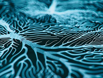
The mechanism that allows these animals to begin the regenerative process involves progenitor cells, connective tissue-associated fibroblasts and skeletal muscle fibers that undergo de-differentiation to become progenitor cells.
Undifferentiated cells usually accumulate at the site of an injury and from there begin the regenerative process. These cell clusters take on properties of cells not associated with their origins and may assume properties of other cell lineages. However, some might also revert to their previous state depending on the specific proteins associated with their origins.
In the axolotl, limb loss results in a buildup of scar tissue, which signals the regenerative process to begin. The presence of transient macrophages allows the signals that promote regeneration until the tissue restoration process is complete, and it appears to happen without incurring genetic abnormalities. This process also appears able to reverse any tumor development by reprogramming abnormal cells. The administration of tumor cells often causes cancer in tissue that is not regenerative, but cancer does not appear when cells are administered into regenerative environments.
There seems to be rudimentary regenerative potential in adult mammals in endogenous stem cells, which are not sufficient to regenerate a functional organ. However, in mammalian embryos, there is a process similar to organ formation in amphibians. Like amphibians, mammalian embryos hold an enormous number of undifferentiated cells that have the potential to have a proper position identity during tissue morphogenesis. These processes are consistent with the behavior of undifferentiated cell clusters in amphibians – the mammalian embryonic environment supports normal tissue development and it possibly has evolved to preclude or minimize malignant transformation.
The question remains: What drives the outcome of embryogenesis, tumorigenesis and regeneration in mammals? What must happen to develop or restore tissue, or disrupt it? Neoplastic transformation in adults is often associated with chronic inflammation. Relatedly, inflammation in tumors is not a self-limiting, continuous activation of wound-healing. This inflammation causes scar tissue, which some investigators believe is the cause of anatomical polarity in tissues undergoing regeneration and is potentially the root of tumorigenesis at the site of an injury.
Another possibility is the accumulation of senescent cells, which are a source of pro-inflammatory factors and represent the senescence-associated secretory phenotype (SASP). SASPs are rich with cytokines and chemokines that can induce a wound-healing response. The disappearance of senescent cells has been identified in the immune cells that surround them, which happens in salamanders during regeneration and in mammalian embryos during morphogenesis. These cells are putative macrophages, which can clear dysfunctional foreign cells.
Macrophages also play a role in constructive and detrimental biological processes. They may hide cues behind the uncontrolled tumorigenic conditions seen in adult mammals. Current tissue engineering recruits and localizes macrophages at implant sites to restore tissue function while avoiding scar formation.
Researchers believe anti-inflammatory macrophages are responsible for activating the biological process associated with extracellular matrix (ECM) formation, collagen fibril reorganization, angiogenesis and tissue morphogenesis, which lead to the complete integration of the scaffold within the tissue.
Embryonic macrophages in mammals during early stages of development also can repair skin wounds or cardiac muscle after ischemic injury in a scarless manner. However, macrophage-depleted neonatal mice are unable to complete these repairs.
Researchers performed a modeling exercise to demonstrate the effect of immune mechanisms in controlling tumor initiation and growth in embryonic stem cells versus embryonic fibroblasts. Based on an extensive literature review, they found the spontaneous mutation frequency in mouse embryonic stem cells is about two orders of magnitude smaller than in differentiated cells such as mouse embryonic fibroblasts.
A cellular automaton is an agent-based model that works on a 2D rectangular lattice composed of square elements (side length 20 µm), representing individual cells that evolve through a set of possible states in discrete time steps based on a set of rules. The cells undergo division to form daughter cells and based on a probability of mutation can acquire mutations or repair them along the way.
Over 384 days, from inception to growth up to one year after birth, simulation results demonstrated that despite a much faster proliferation rate, embryonic stem cells and embryonic fibroblasts gather no mutations until gestation and subsequently start to accumulate mutations only after birth.
This observation contrasts with cancer cells in adults by at least three to four orders of magnitude. In this simplified modeling approach, the authors did not correlate cellular mutation load with malignant transformation, but it could still be inferred that the embryonic microenvironment elicits a protective effect capable of maintaining genomic stability and functionality to minimize the risk of malignant transformations.
Based on the model, the authors conclude that during morphogenesis, the correct patterning for tissue growth is ensured by the precise recapitulation of specific developmental steps along with the immature status of the immune system and the interplay with neighboring cells.
A comparison of the immunological relationship between embryo and tumor with that between embryo and the initial cluster of undifferentiated cells at the regeneration site in regenerating amphibians raises apparent paradoxes. If the investigation of these open questions can be formalized into an experimental hypothesis-generating model, researchers may be able to identify mechanisms that are capable of inducing tissue development in contrast to supporting tumor progression.
Such an approach, based on comparative evolutionary biology, could yield effective regenerative medicine-based strategies to prevent and fight human cancer.
Bruna Corradetti, Prashant Dogra, Simone Pisano, Zhihui Wang, Mauro Ferrari, Shu-hsia Chen, Richard L. Sidman, Renata Pasqualini, Wadih Arap and Vittorio Cristini. Amphibian regeneration and mammalian cancer: Similaritiesand contrasts from an evolutionary biology perspective. February 23, 2021. BioEssays. DOI: 10.1002/bies.202000339.
Erin Graham, January 2022
Share this story
Related Articles
Clinical Research
Cancer cell type (seed) and tumor microenvironment (soil) control therapeutic antibody delivery and efficacy
Primary tumor cells (seed) and metastatic organs (soil) determine complex tumor microenvironment heterogeneities- a major cause of resistance against immunotherapeutics, including immune checkpoint inhibitors important in breast cancer treatment strategies.
Clinical Research
Semantic dementia study yields new findings
Inflammation research uncovers likely therapeutic target for neurodegenerative aphasia disorder